Crucial ECMO Life Support Guide: Advanced Heart & Lung Care
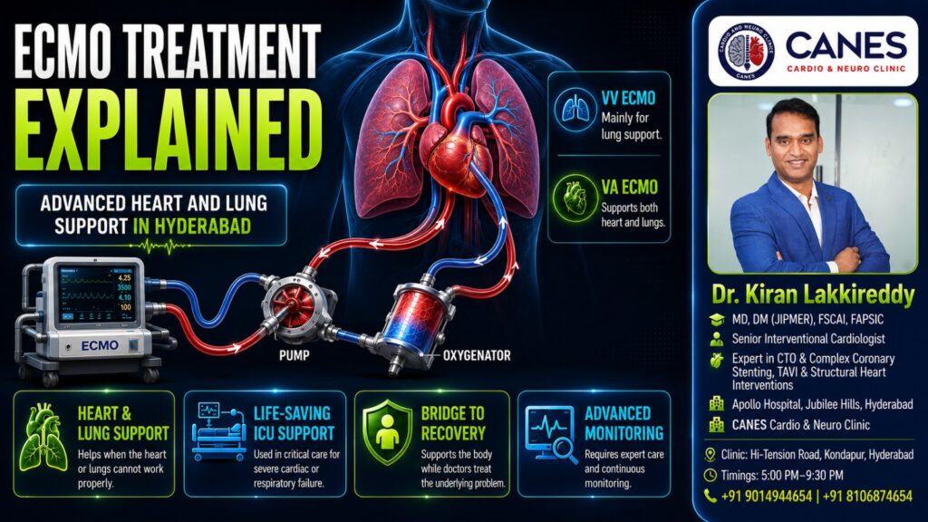
● Lifestyle & Prevention ECMO: Understanding Advanced Heart and Lung Life Support Table of Contents Introduction What Is ECMO? How Does an ECMO Machine Work? What Is the Difference Between VA-ECMO and VV-ECMO? Who May Not Benefit From ECMO? Decision Support: Is ECMO a Last-Resort Treatment? How Long Can a Patient Remain on ECMO? What Are the Possible Risks of ECMO? Myth vs. Fact: Understanding ECMO What Should You Do Next? (A Checklist for Families) Advanced Cardiovascular Evaluation at CANES Clinic FAQs Book a Consultation Prevention is better than cure. Talk to our specialists today. Book Now Severe heart or lung failure can prevent the body from receiving enough oxygen, even when doctors use maximum medical therapy, mechanical ventilation, and other intensive care treatments. In carefully selected, critically ill patients, ECMO life support may temporarily perform part of the work of the heart, lungs, or both organs. ECMO stands for Extracorporeal Membrane Oxygenation. “Extracorporeal” means outside the body. During treatment, blood is removed through large tubes called cannulas, circulated through an external machine, supplied with oxygen, cleared of carbon dioxide, and safely returned to the patient. It is crucial to understand that ECMO does not cure heart or lung disease. Its exact purpose is to maintain circulation and oxygen delivery while the medical team treats the underlying condition, evaluates recovery, or prepares the patient for another life-saving intervention. At CANES Cardio & Neuro Clinic, a specialized cardiovascular care center in Hyderabad, our approach emphasizes clear patient education, careful cardiovascular assessment, and shared decision-making. Guided by Dr. Kiran Lakkireddy, an experienced interventional cardiologist specializing in complex cardiology, we help patients and families navigate serious cardiac conditions and determine when advanced hospital-based care is required. Quick Answer ECMO life support is a temporary, highly advanced system that circulates blood outside the body, adds oxygen, removes carbon dioxide, and returns the blood to the patient. There are two principal forms: VA-ECMO (which supports both the heart and lungs) and VV-ECMO (which supports the lungs but does not directly pump for the heart). ECMO is utilized when potentially reversible heart or lung failure remains life-threatening despite maximum conventional treatments. ECMO Life Support at a Glance Feature What It Means Why It Matters Extracorporeal Circulation Blood travels temporarily outside the body through an external circuit. The machine can oxygenate the blood and, in VA-ECMO, heavily support circulation. Membrane Oxygenator A component that adds oxygen and removes carbon dioxide. It performs part of the gas-exchange work normally carried out by the natural lungs. VA-ECMO / VV-ECMO Artery/Vein configurations for returning blood to the body. VA supports both the heart and lungs; VV supports severe lung failure only. Temporary Support ECMO is usually continued for days or weeks rather than permanently. It provides essential time for recovery, transplantation assessment, or definitive surgery. What Is ECMO? Extracorporeal membrane oxygenation (ECMO) is an advanced form of temporary cardiopulmonary life support used when the heart, lungs, or both organs completely fail to meet the body’s metabolic needs. Key Takeaway: ECMO supports the patient while another treatment or the body’s natural healing process addresses the underlying disease; it is a bridge to recovery, not a cure in itself. Detailed Explanation The ECMO circuit acts in some ways like a modified heart-lung bypass system. However, unlike the bypass machine used for a limited period during open-heart surgery, ECMO may remain in continuous use for days or weeks under strict intensive-care monitoring. Depending on the specific configuration, ECMO life support can add oxygen to the blood, remove carbon dioxide, maintain blood flow to vital organs, and vastly reduce the workload placed on injured lungs. When the heart is severely weakened, it can support circulation to keep the patient alive, acting as a crucial bridge to a ventricular assist device (VAD), transplantation, or natural recovery. How Does an ECMO Machine Work? An ECMO machine creates a controlled external circuit where oxygen-depleted blood is continuously drained through large cannulas, pumped through a membrane oxygenator for gas exchange, warmed, and safely returned to the patient. Key Takeaway: The machine artificially replicates the most vital functions of your heart and lungs, requiring continuous, meticulous calibration by a specialized intensive care team. Detailed Explanation The process relies on a highly sophisticated, continuous loop. First, one or more large tubes called cannulas are placed in major blood vessels in the groin or neck (Cannulation). Low-oxygen blood is drawn from the venous circulation into the ECMO circuit. A mechanical centrifugal pump then moves this blood through a membrane oxygenator, often described as an artificial lung. Here, oxygen enters the blood across a membrane while carbon dioxide is extracted. A heat exchanger warms the blood back to an appropriate body temperature before it is forcefully returned to the patient’s body either into a vein (VV-ECMO) or an artery (VA-ECMO). The circuit is monitored continuously because any changes in pressure, flow, oxygenation, or the appearance of clots require an immediate, expert response. What Is the Difference Between VA-ECMO and VV-ECMO? Venoarterial (VA) ECMO removes blood from a vein and returns it to an artery to support both the heart and lungs, whereas Venovenous (VV) ECMO removes and returns blood solely to the venous system to support only the lungs. Key Takeaway: If a patient’s heart is failing, they require VA-ECMO. If their heart pumps fine but their lungs are failing, they require VV-ECMO. Detailed Explanation In VA-ECMO, because the oxygenated blood is returned to the arterial circulation under mechanical flow, the machine takes over the pumping action of the heart. It is considered for severe cardiac failure (like cardiogenic shock), particularly when circulation remains inadequate despite strong medicines and IV fluids. In VV-ECMO, the patient’s own heart must still pump the freshly oxygenated blood throughout the body. Therefore, VV-ECMO supports severe, acute respiratory failure (such as severe pneumonia or ARDS) but does not directly provide the same circulatory assistance or blood pressure support as VA-ECMO. Comparing ECMO Configurations Consideration VA-ECMO VV-ECMO Primary Purpose Heart and lung support Lung support only Blood
Proven Left Main and Bifurcation Stenting Guide
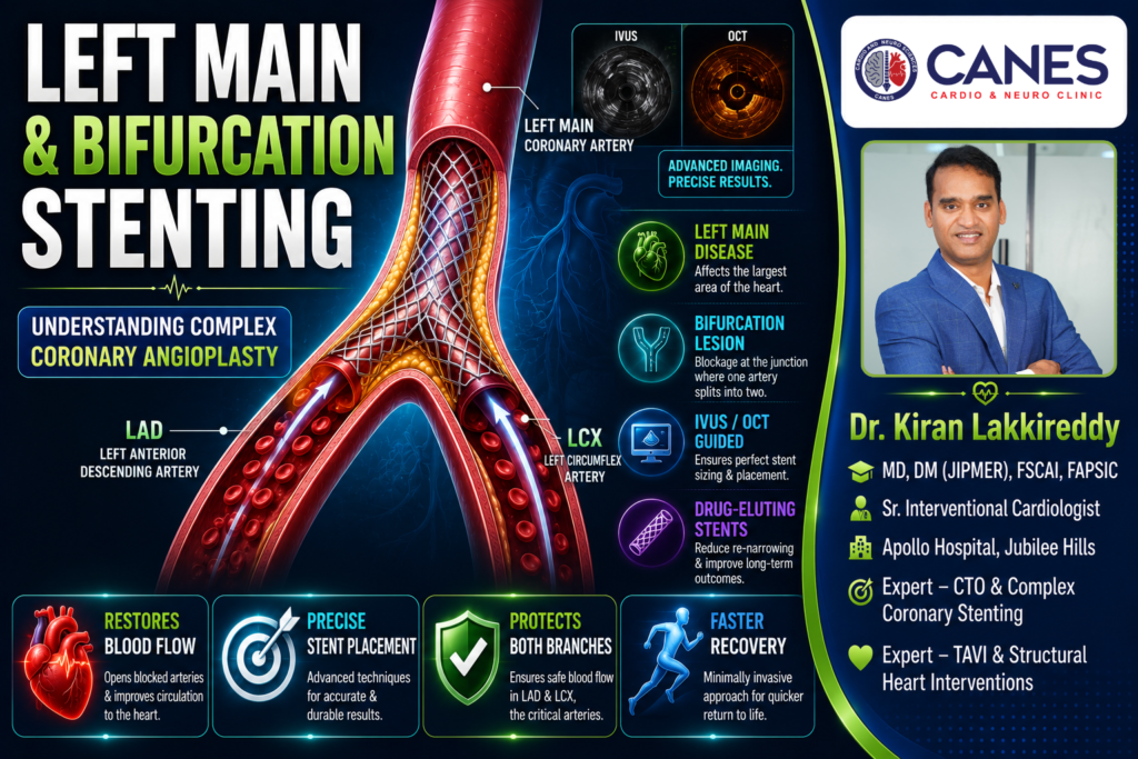
● Lifestyle & Prevention Left Main and Bifurcation Stenting: Understanding Complex Coronary Angioplasty Table of Contents Introduction Complex Stenting At a Glance What Is the Left Main Coronary Artery? What Is a Coronary Bifurcation Blockage? Why Are Left Main and Bifurcation Procedures Considered Complex? Decision Support: Should I Choose Left Main Stenting or Bypass Surgery? How Is Left Main and Bifurcation Stenting Performed? Is One Stent or Two Stents Better for a Bifurcation? Why Is IVUS Important in Left Main Stenting? Who May Not Benefit From Left Main Stenting? What Are the Possible Risks of Left Main and Bifurcation PCI? Myth vs. Fact: Complex Stenting What Should You Do Next? Advanced Cardiovascular Care at CANES Clinic Key Points to Remember FAQs Book a Consultation Prevention is better than cure. Talk to our specialists today. Book Now Coronary stenting is commonly used to open narrowed heart arteries and restore healthy blood flow. However, not every blockage is straightforward. When blockages occur in critical junctions of the heart, specialized techniques like left main and bifurcation stenting become necessary to protect your cardiovascular system. Treating these high-risk locations requires meticulous planning because the cardiologist must open the blocked artery while simultaneously protecting blood flow to every other important branch. Modern drug-eluting stents, advanced imaging, and specialized surgical techniques have made minimally invasive treatment highly successful for carefully selected patients. However, stenting is not automatically the right option for everyone; the decision must be tailored to your specific anatomy, overall health, and long-term goals. At CANES Cardio & Neuro Clinic, a specialized cardiovascular care center in Hyderabad, we believe that patients should clearly understand both their disease and their available treatment options. Guided by Dr. Kiran Lakkireddy, an experienced interventional cardiologist specializing in complex cardiology, our approach focuses on detailed assessment, shared decision-making, and structured follow-up care. In this guide, we explain exactly what left main and bifurcation stenting involves and how experts decide between placing a stent and performing bypass surgery. Quick Answer Left main and bifurcation stenting is a specialized form of coronary angioplasty used to safely treat blockages located in the heart’s primary artery or at an intersection where a vessel divides into two branches. The procedure uses advanced imaging and specialized stent placements to restore essential blood flow. Because this affects a massive area of the heart, treatment decisions are highly individualized, weighing the benefits of minimally invasive stenting against traditional bypass surgery. Complex Stenting At a Glance Feature What It Means Why It Matters Left Main Artery The primary vessel supplying the left side of the heart. A blockage here threatens a massive portion of the heart muscle. Bifurcation A “fork in the road” where one artery splits into two. Treating one branch risks accidentally shifting plaque into the other. IVUS / OCT Microscopic cameras placed inside the artery. Ensures the stent is perfectly sized and expanded for long-term safety. Provisional Stenting Placing one stent first, and a second only if needed. The safest, most common approach to keep procedures as simple as possible. What Is the Left Main Coronary Artery? The left main coronary artery is the major blood vessel beginning at the aorta that divides into the left anterior descending artery (LAD) and the left circumflex artery (LCx), supplying blood to the largest portion of the heart muscle. Key Takeaway: A left main blockage is considered extremely critical because it can threaten the blood supply to a massive area of the heart all at once, unlike a blockage in a smaller branch. Detailed Explanation Left main disease may develop at the very beginning of the artery, in its middle section, or near the intersection where it divides into the LAD and LCx. A blockage at the far end (distal end) of the left main artery is often the most complex to treat because it directly involves both branching vessels. The cardiologist must restore blood flow through the main pipe while avoiding the accidental obstruction of either the LAD or the LCx. While a standard angiogram provides a basic outline of the coronary artery, estimating the true severity of some left main blockages from an X-ray alone can be difficult. For uncertain narrowings, cardiologists rely on advanced intravascular imaging or pressure-based assessments to determine exactly how severely blood flow is being restricted. What Is a Coronary Bifurcation Blockage? A coronary bifurcation blockage is a buildup of cholesterol plaque at a junction where one main blood vessel divides into two distinct branches, creating a Y-shaped intersection. Key Takeaway: Bifurcation stenting is highly complex because inflating a balloon or opening a stent in one branch can physically push plaque into the neighboring branch, accidentally cutting off its blood supply. Detailed Explanation Imagine a major highway that divides into two important exit routes. If a road crew repairs the main highway without protecting the second route, they might accidentally push debris into the exit, closing it off completely. A similar challenge occurs during bifurcation angioplasty. When a balloon or stent expands inside the main vessel, plaque can shift (often called the “snowplow effect”) toward the side branch. The metal structure of the stent itself may also cover the opening of that branch. To prevent this, the cardiologist meticulously plans how to maintain access to both branches, select the exact right stent diameter, and confirm that blood flows freely down both “roads” before finishing the procedure. Why Are Left Main and Bifurcation Procedures Considered Complex? These procedures are considered complex because they involve treating large arteries of varying sizes, navigating challenging vessel angles, and simultaneously preserving high-volume blood flow through multiple critical pathways. Key Takeaway: Procedural success relies not just on placing a stent, but on selecting the correct architectural strategy and using advanced imaging to confirm the stent is structurally perfect. Detailed Explanation Standard stenting is often straightforward, but left main and bifurcation stenting introduces several high-stakes variables: Large Muscle Area at Risk: Left main disease dictates the blood supply for both the LAD and LCx. A mistake here affects